


Please enter the answer below before you can view the full text.
6-1=
Quantitative phase imaging (QPI), promoted by advances in digital holography and computational imaging, is a label-free microscopy approach to investigating phase objects that are optically transparent or translucent, such as cells, tissues, or industrial microstructures. As a computational imaging technique, Fourier ptychographic microscopy (FPM) has rapidly emerged as a powerful tool to achieve wide-field and high-resolution QPI. It can recover the quantitative phase maps of optical path length delays introduced by unlabeled samples, providing an objective measure of refractive index distribution or surface topography of samples. Over the past 12 years, ongoing advances in FPM hardware and software have led to numerous applications in biomedicine and industrial fields. Here, we review the field of QPI based on FPM (FPM-QPI), starting with the underlying principles, followed by a summary of representative advances in both algorithms and imaging models, and ending with the breadth of applications. Finally, we conclude with the emerging challenges and opportunities for the deployment of FPM-QPI as well as the application trends that can expand the scope and utility even further.
Three-dimensional (3D) integral imaging (InIm) is an active research topic in underwater optical imaging and sensing systems. 3D InIm is a passive method to visualize the 3D information of a scene. This method is attractive for imaging in degraded environments and can benefit short to long range applications. In this paper, we present an overview of previously published works on underwater optical imaging and sensing systems showcasing improved visualization and detection capabilities in turbid water using 3D InIm. A set of image sensors capturing images or video sequences were digitally reconstructed using a multidimensional InIm computational reconstruction algorithm for tasks such as visualizing the 3D information of a scene at a particular depth, signal detection, or decreasing the effect of turbidity and partial occlusion. 3D InIm sensing and dedicated computational approaches such as statistical image processing, correlation-based filtering, neural networks, and deep learning make it possible to improve the recovery of information and detection of scenes degraded by turbidity and partial occlusion.
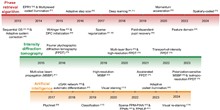
Photoacoustic imaging (PAI) is a non-invasive imaging technique that combines the principles of optical and ultrasound imaging to visualize internal biological structures at high spatial resolution. The major characteristics of PAI, such as its sensitivity to optically absorptive targets (e.g., hemoglobin and melanin), centimeters-deep imaging depth, and non-invasiveness, make it particularly effective in the diagnosis of skin diseases including cancers, inflammatory diseases, and vascular abnormalities. In this review, we categorize PAI modalities according to various imaging scales, i.e., photoacoustic microscopy (PAM), photoacoustic mesoscopy (PAMes), and photoacoustic tomography (PAT), and then discuss their applications for clinical skin imaging in vivo. These modalities provide clinical indicators that quantitatively describe skin vasculature and pigmentation over various spatial resolutions and scanning ranges. We then comparatively discuss and provide insights on the clinical applications of each PAI modality for diagnosing or monitoring various skin diseases.
Fluorescence microscopy technology is a crucial tool in biomedical research, enabling us to visualize the structure and function of tissue cells at the cellular level. Its great potential lies in fluorescence super-resolution fluorescence microscopy, which surpasses the limitations of diffraction and enables high-resolution real-time imaging of nano-subcellular structures, including organelles and cell matrices. It, therefore, contributes significantly to exploring disease mechanisms, ranging from the exchange of protein aggregates at a structural level to the identification of morphological defects in organelles. In this review, we first provide an overview of the principles of various super-resolution microscopy techniques. We then delve into the intricate transmembrane interactions exhibited by membrane organelles, as well as the intricate information communication within membraneless organelles in both physiological and pathological conditions. Finally, we examine the evolution of super-resolution technology and discuss its application in biomedicine. Overall, this review briefly introduces the principles of super-resolution microscopy and highlights its novel results in organelles to guide its application in the biomedical field.
Polarimetric imaging, leveraging measurements of polarimetric parameters that encode distinct physical properties, finds wide applications across diverse domains. However, some critical polarization information is highly sensitive to noise, and denoising polarimetric images while preserving polarization information remains a challenge. The development of denoising techniques for polarized images can be roughly divided into three stages: The first stage involves the direct application of traditional image denoising algorithms, such as spatial/transform domain filtering. The second stage involves specially designed methods for polarized images, using image prior models for noise removal, such as principal component analysis and K-singular value decomposition. In the third stage, benefiting from advances in deep learning, denoising methods tend to integrate polarization characteristics with deep learning models for noise suppression. The residual dense network, U-Net, and other effective models are appropriately modified and supervised/self-supervised trained to handle the denoising problem of regular/extensive polarimetric images. In this paper, we perform a comparative study of polarimetric image denoising methods. These methods are first classified as learning-based and traditional methods. Then, the motivations and principles of different types of denoising methods are analyzed. Finally, some potential challenges and directions for future research are pointed out.
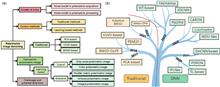
Computational imaging overcomes traditional optical imaging limitations by incorporating encoding and decoding. It represents a paradigm shift in imaging technology, leveraging the manipulation and interpretation of the light field to extract richer information than that was previously attainable. This review explores the emergence and development history of computational imaging. By analyzing the essence of computational imaging from the perspective of the light field, this review categorizes the entire technological roadmap of the computational imaging field based on the imaging framework. This review can serve as a reference for researchers, producers, and policymakers on the main trends, frontiers, and future directions of computational imaging.